
What Is It? A Minimally Invasive Chest Diagnostic Procedure
When I faced cancer, mediastinoscopy was a game-changer. This procedure lets doctors see inside the chest cavity and check for issues.
A mediastinoscopy involves using a thin, flexible tube with a camera to examine the area between the lungs called the mediastinum.
As a two-time lymphoma survivor, I know how vital early detection can be. Mediastinoscopy helps doctors spot cancer in lymph nodes or other chest structures. It’s like having a scout on your team, searching for any signs of trouble.
For men battling cancer, this procedure can feel like a crucial play in the fight. It might seem scary, but knowledge is power. Mediastinoscopy gives us and our doctors the info we need to make intelligent choices about treatment.
Key Takeaways
- Mediastinoscopy examines the chest area between the lungs
- The procedure helps diagnose cancer and other chest conditions
- Recovery is quick, with most patients going home the same day
Understanding Mediastinoscopy
Mediastinoscopy changed my life as a cancer survivor. This procedure helps doctors look inside the chest and diagnose issues like lymphoma.
Purpose and Indications
Doctors use mediastinoscopy to examine the area between the lungs. They insert a thin tube with a camera through a small cut at the base of the neck, allowing them to see lymph nodes and other tissues.
I’ve had this done to check if my cancer spread. It’s also used to diagnose:
• Lung cancer
• Lymphoma
• Sarcoidosis
The procedure can find infections, too. Doctors take tissue samples during mediastinoscopy. These samples help determine the right treatment plan.
For men facing cancer, this test provides crucial info. It guides treatment choices and can improve outcomes.
Historical Perspective
Mediastinoscopy has come a long way since its development in the 1950s. Swedish doctor Eric Carlens developed the technique, which quickly became a key tool for diagnosing chest problems.
Before, doctors had limited ways to see inside the chest. Surgery was often needed, which meant more risk. Mediastinoscopy changed that. It gave a clear view with less invasion.
Over time, the tools improved. Cameras became smaller and clearer, making the procedure safer and more effective.
Today, some centers use video-assisted methods. These give even better views. The basic idea remains the same, though. It’s still about getting a good look at those hard-to-reach areas.
Preparation for the Procedure
Preparing for a mediastinoscopy involves several steps to ensure the best possible outcome. I’ll share my experience to help guide you through this process.
Consultation with Healthcare Provider
When I met with my doctor before my mediastinoscopy, we discussed my medical history and current medications. They explained the procedure in detail, including potential risks and benefits. I asked questions about recovery time and pain management. We also discussed fasting requirements – typically, no food or drink for 8-12 hours before surgery.
My doctor gave me specific instructions on which medications to stop or continue taking. For example, blood thinners often need to be paused. I made sure to mention any allergies, especially to anesthesia or latex.
Pre-Operative Testing
Before my mediastinoscopy, I underwent several tests to assess my overall health. These included:
- Blood tests to check for infection, anemia, and clotting factors
- Chest X-ray to examine my lungs and heart
- CT scan for detailed images of my chest area
- Pulmonary function test to evaluate my lung capacity
I met with the anesthesiologist to discuss my anesthesia options and any concerns. They reviewed my test results and medical history to create a personalized anesthesia plan.
On the day before surgery, I followed all preparation instructions carefully. This included showering with a special soap and avoiding certain foods or drinks. I packed a small bag with essentials for my hospital stay.
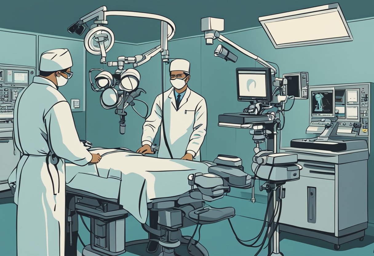
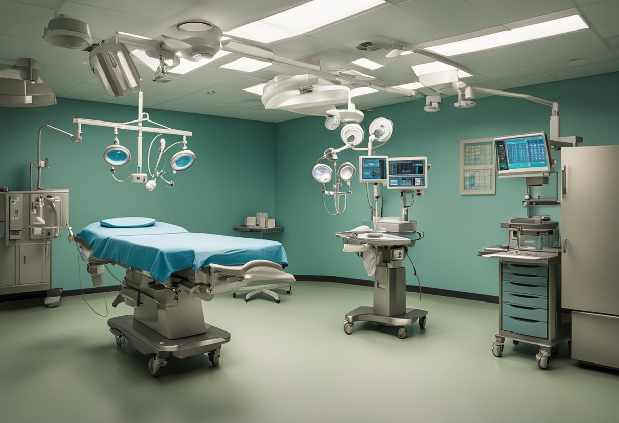
The Mediastinoscopy Procedure
Mediastinoscopy is a surgical technique that lets doctors examine the chest area between the lungs. It’s used to diagnose and stage lung cancer, as well as other conditions affecting this region.
Steps of Mediastinoscopy
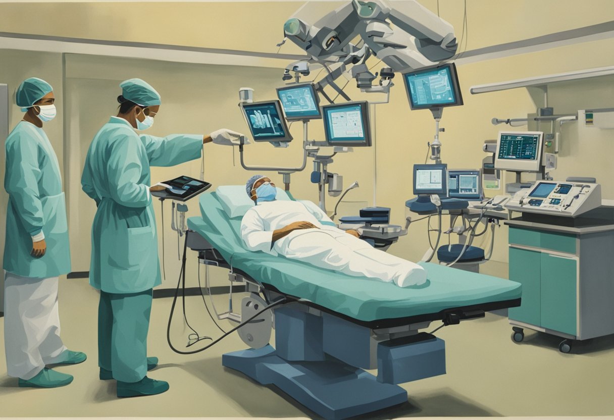
The procedure begins with the patient lying on their back. A small incision is made at the base of the neck, usually about 2-3 centimeters long. The surgeon inserts a thin, flexible tube called a mediastinoscope through this opening. This tool has a light and camera attached, allowing the doctor to see inside the chest cavity.
As a two-time lymphoma survivor, I can attest to the precision of this technique. The surgeon carefully navigates the mediastinoscope to examine lymph nodes and other tissues. They may take small samples (biopsies) for further testing. This step is crucial for accurate diagnosis and staging.
The entire process typically takes about an hour. Once complete, the incision is closed with a few stitches.
Role of Anesthesia
General anesthesia plays a crucial role in mediastinoscopy. It ensures the patient remains still and pain-free throughout the procedure. Before surgery, an anesthesiologist administers medications to induce sleep.
A breathing tube is inserted to help maintain proper oxygen levels during the operation. This tube also prevents any bleeding or fluids from entering the lungs.
As someone who’s been through this, I can say the anesthesia team’s expertise is invaluable. They monitor vital signs and adjust medication levels as needed, ensuring patient safety throughout the procedure.
Potential Risks and Complications
Mediastinoscopy carries some risks and possible complications. As a two-time lymphoma survivor, I’ve learned that understanding these can help patients prepare and recover more smoothly.
Common Complications
Infection at the incision site can occur after a mediastinoscopy. I always remind my fellow cancer warriors to watch for signs like redness, swelling, or fever.
Bleeding is another potential issue. While usually minor, it’s something to be aware of. I’ve found that staying calm and following post-op instructions helps minimize this risk.
Pneumothorax, or collapsed lung, can happen during the procedure. It’s scary but treatable. My experience taught me to communicate any breathing difficulties to the medical team immediately.
Hoarseness might occur if the nerves controlling the vocal cords are affected. As a sports enthusiast, resting my voice, like after a big game, helped speed up recovery.
Risk Factors and Prevention
Certain factors can increase complication risks. Smoking, for example, can affect lung health and healing. I quit before my procedures and felt it made a difference.
Pre-existing heart or lung conditions may also raise risks. It is crucial to be upfront with your doctor about your medical history. I learned this firsthand, and it helped tailor my care.
To reduce risks, follow pre-op instructions carefully. This might include fasting or adjusting medications. Post-op, take it easy like you would after an intense workout. Gradual return to activities worked well for me.
Regular check-ins with your healthcare team are critical. Don’t hesitate to ask questions or voice concerns. Remember, you’re the MVP of your health journey.
What to Expect During Recovery
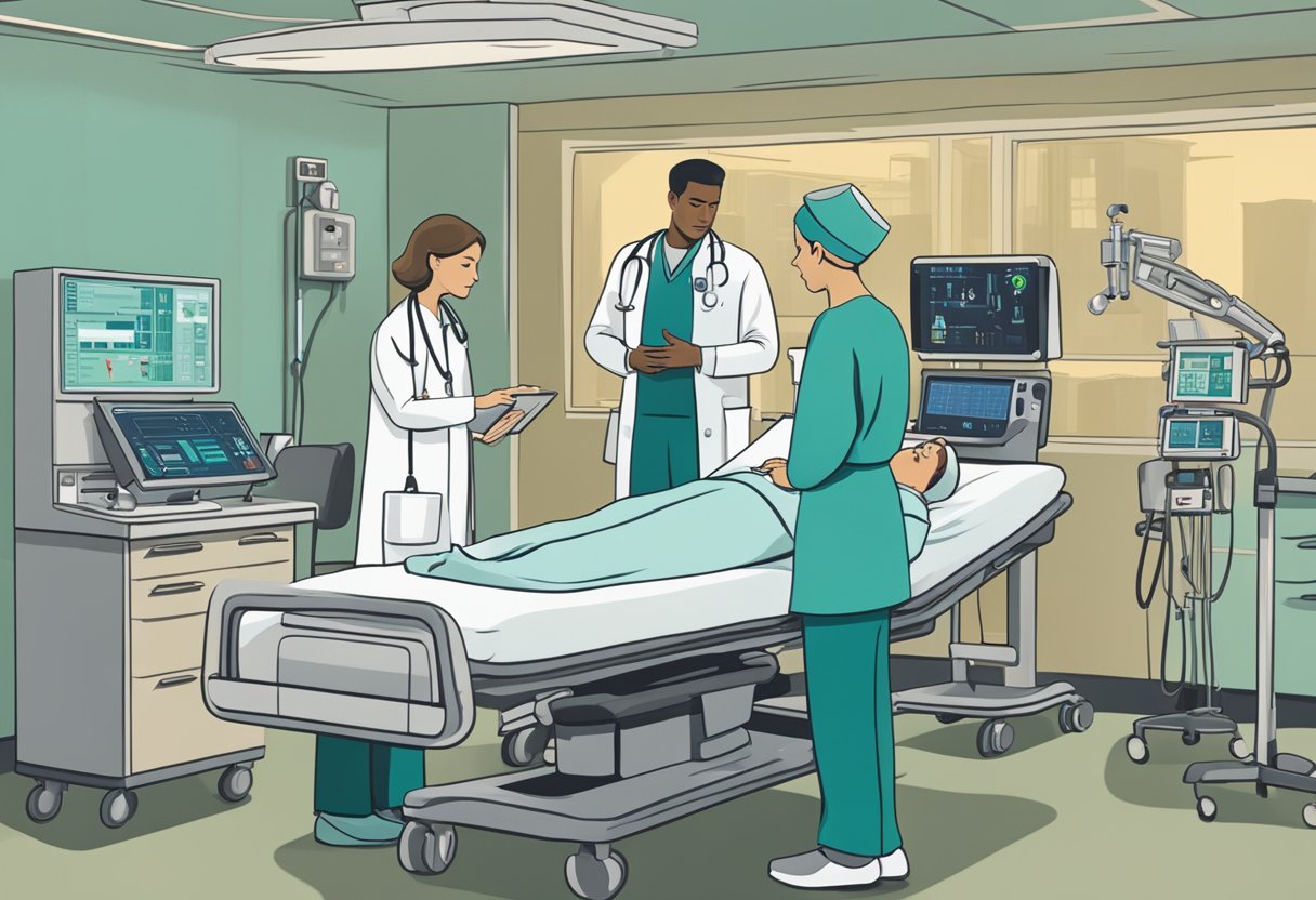
After a mediastinoscopy, your body needs time to heal. The next few days are crucial for a smooth recovery. Let’s explore the critical aspects of post-procedure care and managing side effects.
Post-Procedure Care
I woke up groggy after my mediastinoscopy, feeling like I’d just finished a challenging workout. The nurses kept a close eye on me in the recovery room, checking my vitals and making sure I was comfortable.
Once I was alert, I sipped some water. It felt great on my sore throat. The medical team explained what happened during the procedure and gave me care instructions.
Before heading home, I made sure to ask about pain medicine. They advised me on how to manage any discomfort and when to take the meds.
I learned that most people go home the same day. It’s an outpatient procedure, so there is no need for an overnight stay unless there are complications.
Managing Side Effects
The first few days after my mediastinoscopy were a bit rocky. I felt some pain around the incision site. It was like a constant reminder of what I’d been through.
The breathing tube they used during the procedure made my throat sore. Sipping cool drinks and eating soft foods helped ease the discomfort.
I noticed some swelling and bruising around my neck. The doctor said this was normal and would fade in time.
At first, breathing deeply felt uncomfortable, but I knew it was crucial for preventing lung issues. I did the breathing exercises they taught me, treating them like my new training routine.
I kept my follow-up appointments. These check-ups allowed me to discuss concerns and ensure my recovery was on track.
Interpreting Mediastinoscopy Results
After a mediastinoscopy, your doctor will explain the findings. The results can reveal if cancer has spread to the lymph nodes or other conditions are present.
Understanding Biopsy Results
When I got my biopsy results, I felt nervous. The samples taken during mediastinoscopy are examined under a microscope. This can show if cancer cells are present in the lymph nodes. Typical results mean no cancer was found. Abnormal results may indicate:
- Cancer spread from the lungs
- Lymphoma
- Thymoma
- Infections
My doctor explained each possibility clearly, using simple terms and answering all my questions. This helped me feel more in control of my situation.
Following Up on Abnormal Findings
If abnormal results come back, your doctor will discuss the next steps. My experience taught me to bring a friend to these appointments. They can help remember details and provide support.
Follow-up may include:
- More tests to confirm diagnosis
- Referral to a specialist
- Treatment planning
I found it helpful to write down questions before my follow-up visit. This ensured I didn’t forget anything. Your healthcare team wants to help you understand your condition and options. Don’t hesitate to ask for clarification if needed.
Advancements and Alternatives to Mediastinoscopy
New technologies and methods have changed how doctors examine the chest area. These changes help men with cancer get better care and more options.
Recent Technological Developments
Endobronchial ultrasound has become a game-changer. It uses sound waves to create detailed images of the chest, letting doctors see areas they couldn’t before. I’ve seen how it can find small tumors that other tests might miss.
Doctors now use thin, flexible tubes with cameras and lights. These help them look inside the lungs and chest without significant cuts. It’s like having a tiny explorer in your body. As someone who’s been through cancer twice, I can say this makes a big difference.
CT scans have also improved greatly. They can now show very small details, which helps doctors find problems early and can save lives.
Comparing Mediastinoscopy with Other Procedures
The Chamberlain procedure is another way to check the chest. It’s good for reaching areas that mediastinoscopy can’t. I’ve talked to men who’ve had both, and they say the Chamberlain procedure was more straightforward for them.
Bronchoscopy is less invasive than mediastinoscopy. It uses a thin tube to look in the airways, which for some men might be enough to diagnose what’s wrong.
CT scans are often used before surgery. They help doctors decide if they need to perform more tests, and sometimes, a CT scan alone can give enough information to make a plan.
These new methods mean fewer men need mediastinoscopy. But it’s still essential when other tests don’t give clear answers. -T
Frequently Asked Questions
Mediastinoscopy can be a game-changer for cancer diagnosis and treatment. As someone who’s been through it twice, I’ll share insights on what to expect during this procedure.
What are the potential complications associated with mediastinoscopy?
Complications are rare, but they can happen. Bleeding and infection are possible, like in any surgery. In my experience, a sore throat and chest pain lasted a few days. Some patients might experience temporary hoarseness.
How does the recovery process for a mediastinoscopy typically unfold?
Recovery is usually quick. I was back home the same day. Most people return to normal activities within a week. Rest and follow your doctor’s instructions. Avoid heavy lifting for a couple of weeks.
Can you describe the mediastinoscopy procedure and what it entails?
The procedure involves a small incision at the base of the neck. A thin tube with a camera called a mediastinoscope, is inserted. This lets doctors examine the area between the lungs and take tissue samples if needed.
In what ways does mediastinoscopy differ from bronchoscopy?
Mediastinoscopy examines the space between the lungs, while bronchoscopy looks inside the airways. Mediastinoscopy requires an incision, but bronchoscopy doesn’t. Both can help diagnose lung conditions, but they focus on different areas.
What type of anesthesia is used during a mediastinoscopy?
General anesthesia is used. You’ll be asleep during the entire procedure. I remember feeling nervous, but the anesthesia team was great at putting me at ease before I drifted off.
How is a biopsy performed during a mediastinoscopy?
If the doctor spots suspicious tissue, they’ll take a small sample. Special instruments passed through the mediastinoscope are used for this. Then, the samples are sent to a lab for analysis.
